THE JOINT POLISH-GERMAN Research project

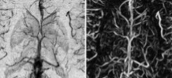
The aim is to develop a MR sequence for high resolution mapping of the cerebral vascular system without the use of contrast agents, as well as segmentation methods of the acquired 3D high resolution images for modeling and quantitative analysis of blood vessel systems. The novel sequence (developed by the Jena team led by prof. J. Reichenbach) will combine time of flight angiography (TOF) and susceptibility weighted imaging (SWI). These two distinct physical effects, flow related enhancement and magnetic susceptibility, are responsible for the contrast of the venous tree and the arterial tree will aid in the endeavor of vessel segmentation, which is the second mile stone of the project. Methods and algorithms of vessel image enhancement and segmentation shall be developed by the research team of Prof. A. Materka in Łódź.
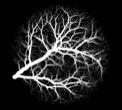
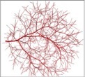
In this joint research project data acquisition as well as post processing in terms of vessel extraction and visualization will be optimized and validated. Vessel tree models extracted from images and image analysis methods will be validated with the use of images simulated and measured for properly designed test objects of known geometry, flow and susceptance parameters of their constituent solid and fluid elements. This will allow assessment of estimation accuracy of extracted parameters of the blood vessels which depends on i.e. image resolution and signal to noise ratio.
The image processing/analysis methods will be implemented in a computer program for veins and arteries visualization and quantitative parameterization, as an aid to medical diagnosis. Potential applications of the project results and methods are manifold. They will not only be supportive to medical diagnosis of brain blood vessel system but also will make a basis for further research on quantitative characterization of vasculature, including thin vessels and capillaries as well as fluids exchange with tissues. The endings of the extracted vessel tree may serve as seed points for mathematical growth models that simulate microvascular architecture, e.g. with the use of 3D image texture analysis and lumped models of nutrients, blood and drug exchange with tissue. Moreover, detailed knowledge of vascular architecture is not only important in surgical planning, but also allows improved characterization of vascular malformations or investigation of the spatial relationship between pathologies and (neo)vascularization.
Project SUMMARY
This research project is supported by the financial mechanism agreed in the Memorandum of Understanding of Scientific Cooperation between The Deutsche Forschungsgemeischaft and The Minister Edukacji i Nauki of the Republic of Poland